- Medicines
- Personal Care
- Skin Cream
- Sunscreen
- Face Wash
- Skin and Body Soap
- Acne Care
- Body Lotions
- Moisturising Lotion
- Moisturising Cream
- Mosquito Repellent
- Moisturising Gel
- Body Wash
- Hair Oils
- Hair Shampoo
- Hair Conditioners
- Hair Supplements
- Hair Colour
- Hair Serum
- Hair Mask
- Hair Solutions
- Baby Diapers and Wipes
- Baby Lotion and Moisturising Cream
- Baby Bath Essentials
- Baby Skin Care
- Baby and Infant Food
- Baby Healthcare
- Women Multivitamins
- Ovulation Test Kit and Women Intimate Care
- Sanitary Pads
- Nutritional Drinks
- Condoms
- Lubricants
- Massage Gels
- Personal Body Massagers
- Men Performance Booster
- Sexual Health Supplements
- Massage Oils
- Ayurveda
- Tooth Paste
- Mouth Ulcer Gel
- Mouthwash
- Toothache and Gum Pain
- Tooth Brush
- Gargle Solution
- Orthopaedic Supports
- Adult Diapers
- Footwear
- Mobility and Support Accessories
- Urinary Support and Care
- Health Conditions
- Bone and Joint Care
- Digestive Care
- Eye Care
- Pain Relief
- Smoking Cessation
- Liver Care
- Stomach Care
- Cold and Cough
- Heart Care
- Kidney Care
- Piles, Fissures & Fistula
- Respiratory Care
- Mental Wellness
- Derma Care
- Pre and Probiotics
- Acidity
- Gas
- Constipation
- Loose Motion/Diarrhoea
- Digestive Fibres
- Digestive Enzymes
- Eye Lubricant Drops
- Lens Solution
- Safety Eye Wear
- Eye Cream
- Eye Vitamins and Supplements
- Eye Drops
- Eye Ointment and Gel
- Nicotine Patch
- Nicotine Gum
- Nicotine Lozenges
- Cough Syrups
- Chest Rubs and Balms
- Nasal Spray
- Lozenges
- Inhalant Capsules
- Cold and Cough Tablets
- Vitamins & Supplements
- Diabetes Care
- Healthcare Devices
- Homeopathic Medicine
- Health Guide
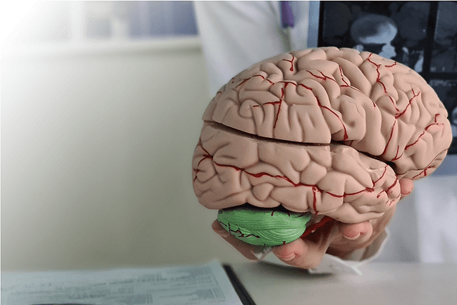
Arteriovenous Malformations
Arteriovenous malformation (AVM) is an abnormal tangle of blood vessels where arteries and veins are directly connected without the presence of capillaries. This condition can occur anywhere in the body but is most commonly found in the brain and spinal cord. Brain AVMs can cause significant neurological symptoms, including seizures, headaches, vision problems, muscle weakness, and in severe cases, hemorrhagic stroke.
Last updated on : 06 May, 2026
Read time : 13 mins
Overview of Disease
An arteriovenous malformation (AVM) is a rare condition characterised by an abnormal tangle of blood vessels in which arteries and veins are directly connected without the presence of capillaries. Capillaries are essential for the normal exchange of oxygen and nutrients between the blood and surrounding tissues. The absence of capillaries in an AVM disrupts the normal blood flow, leading to potential complications such as bleeding, tissue damage, and neurological symptoms. While AVMs can occur anywhere in the body, they are most commonly found in the brain and spinal cord.
What is an Arteriovenous Malformation (AVM)?
Most cerebral AVMs are considered developmental vascular anomalies that are present from early life, although they are often diagnosed later when symptoms arise. Rarely, AVM-like shunts may appear or become apparent later in life; familial syndromes (for example, hereditary haemorrhagic telangiectasia) account for a small proportion of cases. In an AVM, high-pressure arterial blood flows directly into low-pressure veins, bypassing the capillary network. This abnormal connection can cause the blood vessels to become weakened and dilated over time, increasing the risk of rupture and bleeding. The exact cause of AVMs is not well understood, but it is believed to be related to genetic factors and abnormal development of blood vessels during embryonic growth. While AVMs can be a serious condition, it is important to note that many people with AVMs may not experience any symptoms throughout their lives.
Key Factors about Arteriovenous Malformations
| Category | Details |
| Also Referred as | Arteriovenous Malformation (AVM), Cerebral AVM |
| Commonly Occurs In | Usually forms before birth, but symptoms can appear at any age |
| Affected Organ | Brain, occasionally, the spinal cord |
| Type | Abnormal connection between arteries and veins |
| Common Signs | Seizures, headaches, muscle weakness, numbness, vision problems, dizziness, and confusion |
| Consulting Specialist | Neurosurgeon, Neurologist |
| Treatement Procedures | Open brain surgery, embolisation (endovascular treatment), radiation therapy |
| Managed By | Microsurgical resection; endovascular embolisation; Stereotactic radiosurgery; conservative management |
| Mimiciking Condition | Other neurological conditions, such as stroke, brain tumours, or migraines |
Types of Arteriovenous Malformations
Types of AVMs
AVMs can be classified by location (cerebral/intracranial vs spinal vs peripheral), angioarchitecture (compact vs diffuse nidus), and venous drainage patterns (superficial vs deep). For intracranial disease, clinical decision-making is commonly guided by grading systems (for example, the Spetzler–Martin grade and supplemented scores) that combine size, eloquence of adjacent brain, and venous drainage to estimate surgical risk. AVMs are broadly classified by location into intracranial (brain), and spinal AVMs:
1. Brain AVMs
Brain AVMs are the most common type of arteriovenous malformation. They can cause significant neurological symptoms due to their location within the brain tissue. Symptoms can include seizures, headaches, vision problems, muscle weakness, and, in severe cases, haemorrhagic stroke. The severity of symptoms often depends on the size and location of the AVM within the brain.
2. Spinal AVMs
While less common than brain AVMs, spinal AVMs can also lead to serious neurological symptoms. These may include pain, weakness, and paralysis, depending on the location and size of the AVM within the spinal cord. Spinal AVMs can be further classified based on their specific location within the spinal cord and the types of blood vessels involved.
It is essential to recognise the signs and symptoms of AVMs and seek prompt medical attention if an AVM is suspected. Early diagnosis and appropriate treatment can help prevent serious complications and improve patient outcomes.
Symptoms of Arteriovenous Malformations
The symptoms of arteriovenous malformations (AVMs) can vary depending on their location in the brain and whether they have ruptured. Common symptoms include:
- Severe localised headaches
- Seizures occur commonly as a presenting symptom; reported proportions vary in series, but many studies indicate seizures present in roughly 20–40% of symptomatic patients, with haemorrhage and seizures being among the most frequent initial presentations (Chen et al., 2023) .
- Sudden onset of severe symptoms due to a haemorrhage from AVM rupture
- Muscle weakness or paralysis in part of the body
- Vision problems such as blindness or visual field defects
- Confusion and neurological deficits like aphasia, ataxia, and memory problems
- Other symptoms like dizziness, numbness, tingling, pain, and difficulty with movements or speech
In published series, the proportion of AVMs first presenting with haemorrhage varies; many large series report that haemorrhage is the initial presentation in a substantial minority (often around one-third), but exact proportions differ between cohorts (some report ~20–50% depending on referral bias and patient population).
Causes of Arteriovenous Malformations
- Most AVMs are developmental vascular anomalies arising during vascular formation. A small proportion are associated with known genetic syndromes (for example, hereditary haemorrhagic telangiectasia, and the rarer capillary malformation–AVM syndromes). Reports of truly acquired AVMs are rare; the dominant view is developmental rather than de novo adult formation in most cases.
Risk Factors
Several risk factors may increase the likelihood of an AVM causing symptoms:
- Having an AVM in certain areas of the brain prone to bleeding
- Between 15-40 years old, when AVM complications often first appear
- Prior haemorrhage, deep venous drainage, deep brain location, associated aneurysm, and certain angioarchitectural features may increase rupture risk
- Hypertension may worsen outcomes after haemorrhage, but it is not a proven primary cause of AVM rupture
- Sex: Most series report no strong sex predilection; if differences exist, they are small and inconsistent across studies. Age: symptomatic presentation most commonly occurs in adolescents and young to middle-aged adults (commonly cited between ages ~15–40 years).
The specific location and structure of the arteriovenous malformation can affect the risk of rupture and other complications. Age also plays a role, with most AVM symptoms first occurring in people aged 15 to 40. Later, developing AVMs and certain medical issues may also increase the risk (Chen et al., 2023). While these factors can guide screening and treatment, having an AVM does not necessarily mean serious problems will occur.
Prevention of Arteriovenous Malformations
Because most AVMs are developmental anomalies present from early life and there are no established environmental causes, there are no proven primary prevention strategies to prevent AVM formation. Management focuses on early detection of lesions that cause symptoms and individualised treatment planning to reduce haemorrhagic risk and morbidity (Tang et al., 2024).
The Congenital Nature of AVMs and Lack of Prevention
Arteriovenous malformations are intricate vascular lesions that are usually present from birth, although the precise timing of their formation remains unclear. Due to their congenital nature, there are currently no known preventive measures that can be taken to avoid the development of AVMs. Moreover, environmental factors have not been found to play a significant role in their formation, and only a small percentage of cases are linked to genetic syndromes. As a result, the primary focus in managing AVMs is on early detection and appropriate treatment rather than prevention.
Diagnosis & Tests
The diagnosis of an arteriovenous malformation (AVM) typically involves a combination of clinical evaluation and advanced imaging tests. After a thorough interview of the patient's symptoms and a physical examination, doctors use imaging tests to confirm the presence and extent of the AVM.
Initial symptom evaluation often starts with a comprehensive interview to rule out other conditions. Then, various imaging tests are used:
- Computed Tomography (CT) Scan: Uses X-rays to visualise the brain, detect acute bleeding, and show calcifications if present. Less sensitive than MRI for small or non-bleeding AVMs.
- Magnetic Resonance Imaging (MRI): Uses magnetic fields and radio waves to produce detailed images of the brain, assessing the lesion and its surroundings. More sensitive than CT scans.
- Digital subtraction angiography (DSA, catheter cerebral angiography) is the gold standard for defining AVM angioarchitecture (feeding arteries, nidus morphology, and venous drainage) and is essential for detailed treatment planning in most interventional or surgical cases. MRI/MRA and CT/CTA provide noninvasive characterization and are often used for screening or follow-up, but cannot replace DSA when detailed angioarchitecture is required (SNIS Guidelines 2022).
- Ultrasound: In neonates and young infants with open fontanelles, cranial ultrasound or Doppler may identify large vascular lesions; however, ultrasound has limited sensitivity for many intracranial AVMs in older children and adults. The primary imaging modalities for diagnosis and treatment planning are MRI and catheter angiography.
CT scans are useful for detecting acute bleeding and calcifications, but they are less sensitive for small or non-bleeding AVMs. MRI scans provide more detailed images of the brain tissue and are better at differentiating AVMs from other entities like brain tumours. Cerebral angiography is the most detailed test and is crucial for planning treatment as it reveals the exact location of the feeding arteries and draining veins. For young children, ultrasound is a preferred initial test due to its non-invasive nature and lack of need for anaesthesia. Proper diagnosis is crucial for determining the best management and treatment approach for AVMs.
Treatment & Management
The primary therapeutic goals for brain AVMs are to eliminate or reduce the risk of intracranial haemorrhage (when feasible), control symptoms (for example seizures or mass effect), and preserve neurologic function. Management must balance the natural history of the untreated lesion against treatment risks; decisions are individualized and best made by a multidisciplinary cerebrovascular team (SNIS Guidelines 2022). The choice of treatment depends on factors such as the size, location, and symptoms of the AVM, as well as the patient's overall health. Treatment options include:
1. Open Surgery
Microsurgical removal of the AVM is particularly effective for accessible AVMs with low surgical risk, according to the Spetzler-Martin Grade (SMG) scale. The goal is to remove the abnormal tangle of blood vessels completely.
2. Catheter Embolisation
Endovascular embolisation may be used as an adjunct to reduce nidus size or arterial supply before microsurgery or radiosurgery, for staged management of complex AVMs, or as definitive therapy in selected small AVMs or pial fistulas. Its use is highly lesion-specific, and complete cure with embolisation alone is uncommon in many series; combined modality treatment is frequently required.
3. Stereotactic Radiosurgery
Stereotactic radiosurgery (SRS) delivers focused radiation to the nidus and can gradually induce vessel obliteration; obliteration typically occurs over 2–3 years (and in some cases longer), during which the risk of rupture may persist. SRS is most effective for small to medium-sized AVMs and for lesions in deep or eloquent locations where surgery carries high risk.
4. Observation
Conservative management with observation may be chosen for some unruptured AVMs where the estimated lifetime haemorrhage risk is low, and the procedural risk of intervention is higher than the natural history risk. Decisions should be individualised and based on careful risk stratification (size, location, venous drainage, prior haemorrhage, patient age and comorbidity), and patients should be managed in centres with cerebrovascular expertise
5. Symptom Management & Medications
Medical management of complications and supportive care:
- Antiepileptic drugs (AEDs): For seizure control in patients with AVM-associated epilepsy. Choice of AED (for example levetiracetam, sodium valproate/valproate, carbamazepine) should be individualised; levetiracetam is commonly used due to ease of use and tolerability.
- Analgesics: Paracetamol and short courses of nonsteroidal anti-inflammatory drugs (NSAIDs) may be used for headache symptom relief; avoid assumptions that analgesics alter AVM natural history.
- Corticosteroids: May be used short-term to reduce symptomatic cerebral oedema in peri-procedural settings or mass effect; chronic steroid therapy is not a routine treatment for AVMs.
- Blood pressure management: In the acute haemorrhagic setting, blood pressure is managed according to acute intracerebral haemorrhage protocols to limit expansion of haemorrhage. There is no high-quality evidence that long-term use of specific antihypertensive agents prevents AVM rupture; antihypertensives are used to treat hypertension per standard guidelines but are not proven to 'stabilise' AVMs.
- Anticoagulation / antiplatelet therapy: Generally avoided if there is known intracranial AVM because of bleeding risk; any decision to use anticoagulation requires careful multidisciplinary risk assessment (for example when a patient has an unrelated indication such as atrial fibrillation), and may be contraindicated in many cases
When to See a Doctor?
Seek urgent medical attention for sudden severe headache, focal neurological deficits, acute onset of seizure, loss of consciousness, or any signs suggestive of intracranial haemorrhage. For more gradual symptoms such as new-onset seizures or progressive neurologic deficits, prompt evaluation by neurology/neurosurgery is still indicated to allow appropriate imaging and risk stratification. The following key symptoms warrant a prompt medical evaluation:
- Severe headache, which could be a sign of a haemorrhage
- Seizures, as AVMs can disrupt normal brain function and trigger them
- Weakness in the arms or legs, which may result from the mass effect or ischemic injury caused by the AVM
- Vision problems, potentially arising from the AVM affecting areas of the brain responsible for visual processing
- Balance issues, including difficulty with coordination
- Memory and attention problems, which can occur due to abnormal blood flow and potential damage to surrounding brain tissue
Immediate medical care is crucial if any of these symptoms are experienced, as early intervention can significantly impact the outcome and help prevent further complications associated with brain AVMs. By seeking timely medical attention, individuals can ensure that they receive the appropriate diagnosis and treatment, ultimately improving their prognosis and quality of life.
Key Takeaways
- Most brain AVMs are developmental vascular anomalies without proven environmental causes, and there are no established primary prevention strategies; management focuses on early detection, individualised risk assessment, and appropriate treatment when indicated.
- The exact cause of AVMs is unknown, but they are likely related to genetic factors and abnormal blood vessel development in the womb.
- Early detection and proper management are crucial for minimising the risk of complications associated with brain AVMs.
- Regular check-ups and prompt attention to any concerning symptoms can help ensure timely diagnosis and treatment.
- By understanding the nature of brain AVMs and the importance of early intervention, individuals can work closely with their healthcare providers to manage this condition effectively.
FAQs
What are the potential consequences of having a brain AVM?
How long can someone with a brain AVM expect to live?
Is it possible to completely cure a brain AVM?
What are the methods used to remove a brain AVM?
Is recovery possible after being diagnosed with a brain AVM?
Can a brain AVM grow back after treatment?
Is pain a common symptom of a brain AVM?
References
- De Leacy, R., Ansari, S. A., Schirmer, C. M., Cooke, D. L., Prestigiacomo, C. J., Bulsara, K. R., & Hetts, S. W. (2022). Endovascular treatment in the multimodality management of brain arteriovenous malformations: report of the Society of NeuroInterventional Surgery Standards and Guidelines Committee. Journal of NeuroInterventional Surgery, 14(11), 1118–1124. https://doi.org/10.1136/neurintsurg-2021-018632
- Tang, W., Chen, Y., Ma, L., Chen, Y., Yang, B., Li, R., Li, Z., Wu, Y., Wang, X., Guo, X., Zhang, W., Chen, X., Lv, M., Zhao, Y., & Guo, G. (2024). Current perspectives and trends in the treatment of brain arteriovenous malformations: a review and bibliometric analysis. Frontiers in Neurology, 14, 1327915. https://doi.org/10.3389/fneur.2023.1327915
- Chen, Y., et al. (2023). Development and Validation of a Scoring System for Hemorrhage Risk in Brain Arteriovenous Malformations. JAMA Network Open, 2023. (Risk stratification and natural history information).
https://jamanetwork.com/journals/jamanetworkopen/fullarticle/2801834. - Yuan, K., Chen, Y., Yan, D., Li, R., Li, Z., Zhang, H., Wang, K., Han, H., Zhao, Y., Ma, L., Hao, Q., Ye, X., Jin, H., Meng, X., Liu, A., Gao, D., Sun, S., Kang, S., Wang, H., … Zhao, Y. (2024). Re-rupture in ruptured brain arteriovenous malformations: a retrospective cohort study based on a nationwide multicenter prospective registry. Journal of Neurointerventional Surgery, 16(11), 1145–1151. https://doi.org/10.1136/jnis-2023-020650
- Miron, I., Prună, V. M., Visarion, D. M., Petrescu, G. E. D., & Gorgan, R. M. (2024). Natural history and predictors for hemorrhage in supratentorial brain arteriovenous malformations. Journal of Clinical Medicine, 13(13), 3760. https://doi.org/10.3390/jcm13133760
Latest health articles

Top Health Essentials


Disclaimer
Top-Selling Medicines:
...View more
Top-OTC medicines:
...View more
Company
About UsHealth ArticleHealth StoriesHealth LibraryDiseases & Health ConditionsAyurvedaUnderstanding Generic MedicinesAll MedicinesAll BrandsNeed HelpFAQSecuritySavings CalculatorSubscribe
Registered Office Address
Grievance Officer
Download Truemeds
Contact Us
Our customer representative team is available 7 days a week from 9 am - 9 pm.
v4.25.3
2026 - Truemeds | All rights reserved. Our content is for informational purposes only. See additional information.
Our Payment Partners