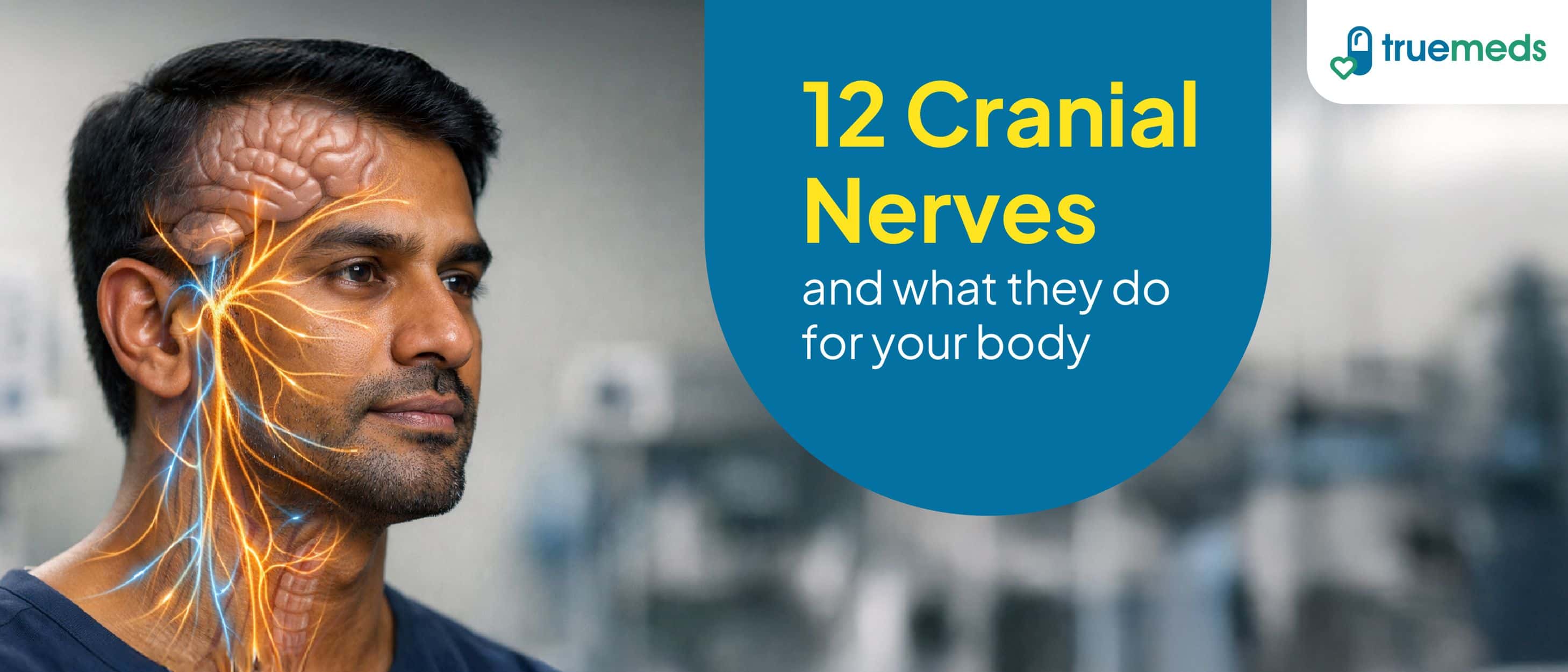
Cranial nerves are essential components of the nervous system, responsible for transmitting sensory and motor information between the brain and the head, neck, and trunk. Twelve pairs of cranial nerves arise from the brain, primarily the brainstem, with exceptions including the olfactory and optic nerves, which originate from the forebrain, and the spinal component of the accessory nerve[1]. These nerves control a wide range of vital functions, including vision, smell, hearing, facial movement, swallowing, and autonomic regulation of heart rate. Additionally, they support both voluntary actions, including eye movement, and involuntary processes[2]. Understanding their anatomy and functions is vital for diagnosing and managing neurological disorders in clinical practice.
What are cranial nerves?
Cranial nerves are specialised peripheral nerves that emerge directly from the brain to innervate structures of the head, neck, and select visceral organs[1]. These nerves are numbered I through XII, starting from the most rostral (closest to the nose) and extending to the most caudal (towards the tail). CN III–XII originate from nuclei in the brainstem, while CN I and CN II are extensions of the forebrain and lack true brainstem nuclei.[3]. Each cranial nerve has its unique sensory, motor, or mixed functions[2]. For example:
- The olfactory nerve (I) transmits smell signals[4]
- The facial nerve (VII) controls facial expressions and taste[2]
- The vagus nerve (X) extends beyond the head and neck, regulating heart, digestive, and respiratory functions[2]
Cranial nerves are categorised by their functions, which can be sensory (e.g., optic nerve for vision), motor (e.g., hypoglossal nerve for tongue movement), or mixed (e.g., trigeminal nerve for facial sensation and chewing)[1]. Their precise function and anatomy make them vital for diagnosing neurological disorders[3].
Did you know? You can test your cranial nerves every day without even realising it! Smelling something fragrant checks your olfactory nerve (CN I), reading uses your optic nerve (CN II), moving your eyes involves CN III, IV, and VI, smiling activates your facial nerve (CN VII), and sticking out your tongue tests your hypoglossal nerve (CN XII).
Overview of the 12 cranial nerves
The cranial nerves are a set of 12 pairs of nerves that originate from the brain and brainstem. They play a vital role in transmitting sensory and motor information between the brain and various parts of the head and neck[1]. Here is an overview of the 12 cranial nerves:
1. Olfactory nerve (CN I)
The olfactory nerve is responsible for the sense of smell. It transmits sensory information from olfactory receptors in the nasal mucosa to the brain. This nerve allows us to detect and differentiate between a wide range of odours, playing a vital role in taste perception and alerting us to environmental dangers such as smoke or spoiled food[4]. It is purely sensory and does not have any motor functions.
2. Optic nerve (CN II)
The optic nerve carries visual information from the retina to the brain's visual cortex[1]. It is made up of nerve fibres from retinal cells that converge at the optic disc and transmit signals that allow us to perceive light, shapes, colours, and movement. Damage to the optic nerve can lead to vision loss or visual field defects[3].
3. Oculomotor nerve (CN III)
The oculomotor nerve controls most of the eye's movements, including upward, downward, and inward motion. It also raises the upper eyelid (via the levator palpebrae superioris) and constricts the pupil for near vision (accommodation)[2]. Dysfunction can result in a drooping eyelid (ptosis), dilated pupil, or double vision due to impaired eye movement[3].
4. Trochlear nerve (CN IV)
The trochlear nerve innervates the superior oblique muscle of the eye, which helps rotate the eye downward and outward[2]. It is the smallest cranial nerve and the only one to emerge from the dorsal side of the brainstem[1]. Damage may lead to difficulty looking down, particularly noticeable when walking downstairs[3].
5. Trigeminal nerve (CN V)
The trigeminal nerve has three branches: ophthalmic, maxillary, and mandibular. The trigeminal nerve provides general sensory input from the face, scalp, sinuses, and anterior two-thirds of the tongue (excluding taste). It also supplies motor fibres to the muscles involved in chewing[2]. Trigeminal neuralgia is a painful condition associated with this nerve and can cause intense facial pain[5].
6. Abducens nerve (CN VI)
The abducens nerve controls the lateral rectus muscle, allowing the eye to move outward (abduction). It is essential for coordinated side-to-side eye movement[2]. Damage to this nerve may result in the inability to move the eye laterally, leading to horizontal double vision[3].
7. Facial nerve (CN VII)
The facial nerve is responsible for facial expressions by innervating the facial muscles. It also carries taste sensations from the front two-thirds of the tongue and provides parasympathetic fibres to the lacrimal (tear) and salivary glands[2]. Bell's palsy, a condition marked by sudden facial weakness or paralysis, often results from facial nerve inflammation[5].
8. Vestibulocochlear nerve (CN VIII)
This nerve has two components: the cochlear nerve for hearing and the vestibular nerve for balance. It transmits sound and equilibrium information from the inner ear to the brain[2]. Damage to this nerve can result in hearing loss, vertigo (a spinning sensation), or balance disorders such as dizziness and difficulty walking or maintaining balance[3].
9. Glossopharyngeal nerve (CN IX)
The glossopharyngeal nerve assists in swallowing and carries taste from the posterior third of the tongue. It also innervates the parotid salivary gland and plays a role in monitoring blood pressure and oxygen levels via the carotid body and sinus[2]. Impairment can affect swallowing, taste, and cardiovascular reflexes[3].
10. Vagus nerve (CN X)
The vagus nerve is the longest cranial nerve and has the most extensive distribution, reaching from the brainstem to the abdomen. It regulates vital autonomic functions, including heart rate, digestion, and respiratory rate. It also innervates the muscles of the voice box (larynx) and soft palate[2]. Vagal nerve damage can cause hoarseness, difficulty swallowing, and digestive issues[3].
11. Accessory nerve (CN XI)
The accessory nerve controls the sternocleidomastoid and trapezius muscles, allowing head rotation and shoulder elevation[2]. It has a spinal component responsible for head and shoulder movement; the cranial portion functionally merges with the vagus nerve.[1]. Injury to this nerve can result in weakness when turning the head or shrugging the shoulders[3].
12. Hypoglossal nerve (CN XII)
The hypoglossal nerve controls tongue movements essential for speech, chewing, and swallowing. It innervates the intrinsic and extrinsic muscles of the tongue[2]. Damage may lead to tongue deviation towards the affected side and speech difficulties[3].
Clinical significance of the cranial nerves
Cranial nerve impairments can have a significant impact on an individual's health and quality of life. These impairments can manifest as sensory deficits, motor dysfunction, or autonomic disruptions, depending on the specific nerve affected[3]. Some common disorders associated with cranial nerve damage include:
- Anosmia (loss of smell) due to olfactory nerve (I) damage[4]
- Vision loss or visual field defects resulting from optic nerve (II) damage[3]
- Trigeminal neuralgia or chewing difficulties caused by trigeminal nerve (V) dysfunction[5]
- Bell's palsy, a unilateral facial drooping and impaired taste, due to facial nerve (VII) palsy[5]
- Hearing loss, vertigo, or balance issues stemming from vestibulocochlear nerve (VIII) damage[3]
- Disruption of heart rate, digestion, or speech as a result of vagus nerve (X) dysfunction[3]
- Swallowing difficulties or tongue movement impairment due to glossopharyngeal (IX) or hypoglossal (XII) nerve injuries[3]
Diagnosing cranial nerve impairments involves targeted examinations, such as olfactory testing with familiar scents, pupil reactivity checks (II, III), facial muscle assessments (VII), and gag reflex evaluation (IX, X)[3]. The gag reflex is inconsistently present in healthy individuals and should be interpreted cautiously. Imaging techniques like MRI or CT scans can help identify structural lesions, whilst electromyography assesses motor nerve integrity[3].
Professional diagnosis is essential. Do not attempt to self-diagnose.
Prompt recognition of symptoms, including ptosis (III), diplopia (IV, VI), or dysphonia (X), is crucial for effectively managing conditions such as stroke, tumours, or neuropathies[3].
Conclusion
The cranial nerves play a vital role in our daily lives, facilitating sensory perception, motor control, and autonomic regulation. A thorough understanding of the 12 cranial nerves and their functions is crucial for doctors to accurately diagnose and effectively treat various neurological disorders. By recognising the symptoms associated with specific cranial nerve impairments, such as vision loss, facial drooping, or swallowing difficulties, doctors can promptly initiate appropriate management strategies. Early detection and intervention are key to improving patient outcomes and minimising the impact of these conditions on an individual's quality of life.
FAQs
What are cranial nerves, and why are they important?
Cranial nerves are twelve pairs of nerves controlling sensory and motor functions of the head, neck, and internal organs. They are essential for vital functions, including vision, hearing, taste, smell, facial movement, and autonomic regulation of the heart and digestive systems.
How can cranial nerve damage affect the body?
Damage to the cranial nerves may cause weakness, paralysis, sensory loss, vision or hearing problems, facial asymmetry, or impaired swallowing and speech. The specific effects depend on which nerve is affected and the severity of the damage.
What are common symptoms of cranial nerve dysfunction?
Symptoms include double vision, facial numbness, muscle weakness, hearing loss, dizziness, taste changes, and difficulty speaking or swallowing. Some individuals may also experience drooping eyelids, dilated pupils, or balance problems.
How are cranial nerve disorders diagnosed?
Doctors use neurological exams, imaging (MRI, CT), nerve conduction studies, and symptom history to identify nerve dysfunction accurately. Specific tests include pupil reactivity, facial muscle assessment, and sensory evaluations.
Can cranial nerve damage be treated or reversed?
Some nerve damage is treatable or reversible; recovery depends on the cause, severity, and promptness of medical intervention. Treatment may include medications, physical therapy, or surgical procedures, depending on the underlying condition.
What are some common cranial nerve disorders?
Examples include Bell's palsy, trigeminal neuralgia, optic neuritis, vestibular neuritis, and cranial nerve palsies from stroke. These conditions can significantly impact daily functioning and quality of life.
How do cranial nerves relate to brain function and health?
They connect the brain to sensory organs and muscles, playing critical roles in daily functions and neurological health. Cranial nerves serve as vital communication pathways, enabling the brain to receive information and control bodily functions.
Medical Disclaimer
This article is for informational purposes only and does not constitute medical advice. The information provided should not be used for diagnosing or treating health conditions. Always consult a qualified healthcare provider for diagnosis, treatment, and personalised medical advice. Do not disregard professional medical advice or delay seeking it because of information found in this article. If you have a medical emergency, contact your doctor or emergency services immediately.
References
Sonne, J., Omole, A. E., & Lopez-Ojeda, W. (2025, January 24). Neuroanatomy, cranial nerve. StatPearls - NCBI Bookshelf. https://www.ncbi.nlm.nih.gov/books/NBK470353/
Porras‐Gallo, M. I., Peña‐Meliáan, Á., Viejo, F., Hernáandez, T., Puelles, E., Echevarria, D., & Sañudo, J. R. (2018). Overview of the history of the cranial nerves: From Galen to the 21st century. The Anatomical Record, 302(3), 381–393. https://doi.org/10.1002/ar.23928
MSD Manuals. (2025). How to assess the cranial nerves. MSD Manual Professional Edition. https://www.msdmanuals.com/professional/neurologic-disorders/neurologic-examination/how-to-assess-the-cranial-nerves
Reese, V., Das, J. M., & Khalili, Y. A. (2023, May 6). Cranial nerve testing. StatPearls - NCBI Bookshelf. https://www.ncbi.nlm.nih.gov/books/NBK585066/